Published On Sep 26, 2014
Phases of mitosis:This animation demonstrates the stages of mitosis in an animal cell.
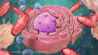
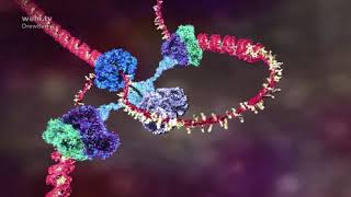
Mitosis is the process in which a eukaryotic cell nucleus splits in two, followed by division of the parent cell into two daughter cells. The word "mitosis" means "threads," and it refers to the threadlike appearance of chromosomes as the cell prepares to divide. Early microscopists were the first to observe these structures, and they also noted the appearance of a specialized network of microtubules during mitosis. These tubules, collectively known as the spindle, extend from structures called centrosomes - with one centrosome located at each of the opposite ends, or poles, of a cell. As mitosis progresses, the microtubules attach to the chromosomes, which have already duplicated their DNA and aligned across the center of the cell. The spindle tubules then shorten and move toward the poles of the cell. As they move, they pull the one copy of each chromosome with them to opposite poles of the cell. This process ensures that each daughter cell will contain one exact copy of the parent cell DNA.
Interphase: Cells may appear inactive during this stage, but they are quite the opposite. This is the longest period of the complete cell cycle during which DNA replicates, the centrioles divide, and proteins are actively produced. For a complete description of the events during Interphase, read about the Cell Cycle.
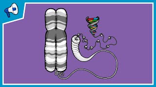
Prophase: During this first mitotic stage, the nucleolus fades and chromatin (replicated DNA and associated proteins) condenses into chromosomes. Each replicated chromosome comprises two chromatids, both with the same genetic information. Microtubules of the cytoskeleton, responsible for cell shape, motility and attachment to other cells during interphase, disassemble. And the building blocks of these microtubules are used to grow the mitotic spindle from the region of the centrosomes.
Prometaphase: In this stage the nuclear envelope breaks down so there is no longer a recognizable nucleus. Some mitotic spindle fibers elongate from the centrosomes and attach to kinetochores, protein bundles at the centromere region on the chromosomes where sister chromatids are joined. Other spindle fibers elongate but instead of attaching to chromosomes, overlap each other at the cell center.
Metaphase: Tension applied by the spindle fibers aligns all chromosomes in one plane at the center of the cell.
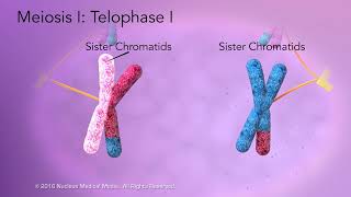
Anaphase: Spindle fibers shorten, the kinetochores separate, and the chromatids (daughter chromosomes) are pulled apart and begin moving to the cell poles.
Telophase: The daughter chromosomes arrive at the poles and the spindle fibers that have pulled them apart disappear.
Cytokinesis: The spindle fibers not attached to chromosomes begin breaking down until only that portion of overlap is left. It is in this region that a contractile ring cleaves the cell into two daughter cells. Microtubules then reorganize into a new cytoskeleton for the return to interphase.
#mitosis #meioses #biology #celldivision #viralvideos #fertilisation